Atrium Health Navicent Gastroenterology
Endoscopic Ultrasound

According to medicinenet.com, Endoscopic Ultrasound is a combination of both endoscopy and ultrasound technologies used to extract images of our digestive tract, surrounding tissues, and other internal organs. Endoscopy refers to the practice of inserting a flexible tube (usually long) through either the rectum or mouth to visualize the digestive tract. Further, ultrasound relates to the method of using high-frequency sound waves to produce images of internal organs, which could include liver, uterus, ovaries, pancreas, and aorta, among other organs.
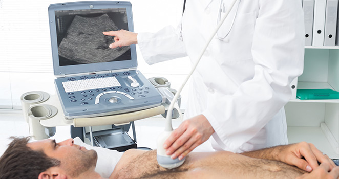
Typically, a basic ultrasound sends sound waves to target organ(s) and receives feedback with a transducer placed on the skin and closer to the body to be imaged. However, it should be noted that images are not of high quality. On the contrary, in Endoscopic Ultrasound, an ultrasound transducer is strategically placed at the tip of the endoscope and inserted into the body. Therefore, by insertion, the device obtains high-quality images of the digestive tract.
The placement of the transducer at the tip of the endoscope is meant to ensure it gets as close as possible to the internal organs. This proximity provides images, which are more detailed and accurate than the traditional ultrasound. The endoscopic ultrasound is also able to show information on the intestinal walls and adjacent areas.
Doppler ultrasound is an application of endoscopic ultrasound used to capture images of blood flow and obtain tissue samples. The tissues may then be used by pathologists under a microscope for any medical investigation they find fit.
When is the Procedure Useful?
The application of Endoscopic Ultrasound is still a developing area, and we cannot exhaustively give a list of its uses in the medical field; more medical possibilities and discoveries could be in the offing. However, currently, these are some of its applications:
- Diagnosing cancers of the pancreas, stomach, rectum, and esophagus.
- Diagnosing lung cancer
- Examining chronic pancreatitis and cysts of the pancreas
- Checking abnormalities of the bile duct or gallbladder.
It is imperative to note that Endoscopic Ultrasound is commonly used in the staging of cancer. If the procedure is carried out on time, it can provide measures to ensure that the problem is averted where possible. For example, early stages colon cancer refers to cancer that is initially confined to the inner colon just before it starts spreading to other parts of the organ. Therefore, if Endoscopic Ultrasound is used to detect cancer early enough, it is possible to reset the condition, and a patient may heal completely.
In cases where cancer is detected at advanced stages, it will have penetrated the colon and affected the adjacent organs. Endoscopic Ultrasound can provide details on the depth of the tumor penetration in the body. This is useful information, which can be used during the staging phase.
Preparation for Endoscopic Ultrasound
Before the Endoscopic Ultrasound procedure is started, the doctor will have to determine if you are a good fit for it or if contraindicated due to allergies. The doctor will also seek to find out if you have other health complications including diabetes mellitus, lung disease, or heart disease. The doctor may also want to determine if you have any bleeding disorders or at risk of bleeding or infection. Full disclosure of personal medical history and family history, which may place you at risk, is important.
Endoscopic Ultrasound is administered with sedation and in most cases; a patient is not able to resume regular duties for up to 24 hours afterward. This procedure is commonly an outpatient procedure, and you will need to be driven or have a ride home. Before the procedure, it is highly recommended you do not eat anything for at least 6 hours. The doctor will give you an order for laxatives and/or enemas prior to a rectal procedure. They encourage you to follow the directions strictly to prevent any delays in your procedure
How Endoscopic Ultrasound is Performed
Once you arrive for the procedure, you will be guided and advised by the doctor on what it will entail, and any concerns will be addressed. Before the procedure is conducted, you will be given a consent form, and you will be expected to sign explicitly stating that you are fully conscious of what it will entail together with the risks associated with it. All necessary preparations will be conducted before you will be taken to the theater where sedation will be administered. Electrode patches to monitor your pulse, blood pressure, or blood oxygen will be placed on your skin.
Once sedation is administered, a patient may feel some slight discomfort if any. The doctor will observe the images of the target organs on a screen displaying them. Depending on complexity, the entire procedure could take about 30 to 90 minutes. Depending how you recover from the procedure, you may be discharged or kept in hospital overnight for close monitoring.
Risks Associated with Endoscopic Ultrasound
An Endoscopic Ultrasound procedure is safe; complications associated with it are rare. However, one of the common side effects is that patients may develop skin rashes, nausea, or hives during treatment. Patients are advised to visit a physician if the problem persists. When needles are used for the Endoscopic Ultrasound, proper care should be taken to ensure that intestinal lining is not affected.
It should be noted that for the procedure to be successful, it takes the input of all stakeholders. Medical practitioners have to conduct due diligence to ascertain if it would be necessary to perform Endoscopic Ultrasound and the possible contemplated outcomes of the procedures. On the other part, patients have to ensure they stick to guidelines provided to them by the doctor for the success of the process.