Atrium Health Navicent Heart & Vascular Care
Vascular Lab
The heart and blood vessels conform to what it is known as the circulatory system. Functions of the heart are to pump blood into any of the three major types of vessels in the body:
- Arteries: Which transport oxygen-rich blood to the tissues and limbs.
- Veins: Which make blood return from tissues and the limbs to the heart for it to be restocked with oxygen by the lungs.
- Lymphatics: Which send fluid from the skin and tissues back to the veins.
It is possible that, over the course of time, veins and arteries in the body start to malfunction and cause sudden symptoms and complications that will affect a person's health. It is necessary then to properly identify the problem through a specific test.
What is a Vascular Lab?
A vascular lab is a non-invasive ultrasound test to help diagnose vein disorders and diseases and address many vascular conditions --such as peripheral arterial disease (PAD), thoracic outlet syndrome, stroke aneurysms, etc. Following a doctor's physical examination, being tested in a vascular laboratory is frequently the starting point for detecting these types of disorders, and once a doctor has confirmed a diagnosis, a course of treatment will be established. In addition to the ultrasound, segmental pressure/volume readings could be taken in order to identify the type of vascular disease.
How does it Work?
These exams are to be performed by a licensed vascular technologist. The standard procedure analyzes blood vessels in the body that are vital for the functioning of major organs and tissue. However, specific exams are to be ordered according to the patient's complaints and suspected vascular conditions.
Non-invasive examining operates numerous types of technology to assess movement, perfusion, and compressions inside the vessels when the body is stationary and exercising. Pulse-volume and segmental pressures recordings are other significant methods used in the vascular laboratory. Both these techniques are largely painless and help define the location and severity of the problem.
Ultrasound equipment is used to take photographs of blood vessels and to evaluate blood flow in these vessels. Some of the latest transportable ultrasound machines get to be the size of a laptop computer and the tiniest ultrasound machines easily fit into a physician's laboratory coat pocket. Nonetheless, these hand-held Doppler devices are not suitable for imaging vessels but for listening to blood flow.
Preparation
With most vascular tests, there are generally no prior dietary restrictions; patients may eat and drink at their discretion. The duration of the exam is usually between 30 to 90 minutes approximately.
However, there might be a few exams that do require special preparation such as the Abdominal Duplex Doppler exam. For this procedure the patient should not eat or drink past midnight the night before; should avoid dairy products, raw vegetables, or any food that may produce gas, the day before since gas pockets have a negative effect on the quality of the pictures and could cause the test to be rescheduled.
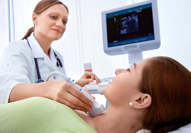
During a Vascular Lab
For the duration of the procedure, sound waves are conducted through the tissues of the area being tested. Then the sound waves reflect off the blood cells moving inside the blood vessels. This method allows the physician to estimate their speed, record it, and get the readings displayed on a screen.
A registered technologist will perform the test and a vascular physician will be in charge of interpreting the readings as the patient is lying on an expanded examining table. A small quantity of water-soluble gel is administered to the skin in the area to be examined. This kind of ointment causes no harm to the skin. Then a small piece of equipment called a transducer is positioned over the testing zone and the images produced by it should be displayed on the ultrasound screen. The transducer remains in place until the computer is able to record the blood flow data.
If instead of an ultrasound, a pulse-volume recording lab is being performed, the registered vascular technologist will place a set of blood pressure cuffs around the patient's legs or arms. These bracelets will then be inflated at various levels in the arm and leg to acquire segmental blood pressures and waveforms using the pulse-volume recording equipment and a Doppler device.
There is nearly no discomfort throughout the process, however, the patient may be able to hear the technologist taking measurements, listening to the blood stream and handling the equipment.
If surgery is required after the test, a special ink may be used to mark veins on the surface of the skin -as a request from the surgeon- so they can be located during the operation. It is important not to wash these marks off. At any rate, the patient may be given a pen to correct these where they are not visible enough.
What Happens After?
The water-soluble solution will be wiped off the patient's skin. No special home-care is needed after the exam. The patient may go home right away.
What are the Side Effects?
This procedure causes no harmful effects on the patient. Furthermore, noninvasive vascular examining does not make use of radiation, which x-rays do.
Knowing the Results
A final report will be produced once a vascular medicine physician checks on the results. This document is then sent out to the patient as a doctor's visit is scheduled to discuss the findings.
What Kind of Tests Can Be Done?
These are some of the studies that can be performed at a vascular laboratory:
- Vein and arterial mapping
- Carotid, renal and peripheral artery ultrasound
- Abdominal aorta ultrasound
- Pulse volume readings
Keep in mind that
Vascular lab tests are also useful in tracking the evolution of a disease over time.